
INTRODUCTION
Alginate, also known as alginic acid, is a natural polysaccharide found in brown algae. It has a linear copolymer structure comprised of homopolymeric blocks of (1→4)-linked β-D-mannuronate and α-L-guluronate residues. Given the similarity of its chemical structure to polysaccharides naturally present in the extracellular matrix of human tissues, as well as its ability to form hydrogels at physiological conditions of pH, temperature, and ionic strength, by simple ionical cross-linking with bivalent cations such as Ca2+ or Mg2+, alginate-based hydrogels are of great interest for the engineering of biological tissues. Especially, these hydrogels have been extensively applied in cartilage tissue engineering, in which histological processing is a must for the evaluation of the quality of the engineered tissue.
Routine histological techniques such as Alcian Blue or Safranin O staining are used for the manifestation of cartilage-specific extracellular matrix (ECM) deposition. These techniques rely on the binding of positively-charged dyes to the negatively-charged polysaccharide chains that are specifically accumulated in the ECM of cartilage; namely hyaluronic acid, chondroitin sulfate and keratan sulfate. These polysaccharide molecules possess negative charges due to the presence of carboxylic acid and or sulfate groups in their chemical structure. In the case of alginate, carboxylic acid groups are present in its chemical structure; which will bind positively-charged dyes upon histological staining. This makes the identification of cartilage-specific ECM deposition in alginate-based hydrogels, technically challenging by means of routine histological techniques.
Protonation of carboxylic acid and sulfate groups at pH values below physiological ranges can neutralize the negative charges that bear these functional groups. As protonation of different functional groups occur at different pH values, this phenomenon can be exploited to selectively neutralize the different functional groups, and therefore, discriminate between strongly sulfated, strongly and weakly sulfated, and sulfated and carboxylated polysaccharides, by means of adjusting the pH of Alcian Blue staining solution to 0.2, 1.0, and 2.5, respectively.
MATERIALS
- Xylene
- 100º Ethanol
- 95º ethanol
- 70º ethanol
- H2O, distilled
- HCl 0.1M, pH 1.0
- Alcian Blue, 1% (w/v) in HCl 0.1M, pH 1.0
- Nuclear Fast Red staining solution, 0.1% (w/v)
- Glass coverslips
- Mounting medium (DPX or similar)
EQUIPMENT
- Chemical fume hood
- Microscope
- Coplin jars
- Tweezers
METHODS
Follow institutional and local guidelines for the safe handling and disposal of chemical reagents. Prepare 95º and 70º ethanol from 100º ethanol and distilled water. Prepare 0.1M HCl pH 1.0 by diluting concentrated HCl in distilled water and adjuting the pH with concentrated HCl. Alcian Blue 1% (w/v) solution in HCl 0.1M (pH 1.0) and Nuclear Fast Red 0.1% (w/v) can be purchased in a ready-to-use format.
Start with formalin-fixed paraffin-embedded histological sections mounted onto microscopy glass slides. For performing the Alcian Blue – Nuclear Fast Red stain, follow the steps listed below:
- Xylene, 10 min
- Xylene, 10 min
- 100º ethanol, 5 min
- 100º ethanol, 5 min
- 95º ethanol, 5 min
- 70º ethanol, 5 min
- H2O distilled, 5 min
- HCl 0.1 M pH 1.0, 2 min
- Alcian Blue, 1% (w/v) in HCl 0.1M, pH 1.0, 30 min
- HCl 0.1 M pH 1.0, 2 min
- Nuclear Fast Red staining solution 0.1% (w/v), 5 min
- H2O distilled, 2 min
- 70º ethanol, 2 min
- 95º ethanol, 3 min
- 100º ethanol, 3 min
- 100º ethanol, 3min
- Xylene, 5 min
- Xylene, 5 min
- Mount glass coverslips using DPX or similar
- Leave set mounted histological preparations overnight in horizontal position
- Visualize under brightfield microscopy
RESULTS
Following the protocol described above, selective staining of cartilage-specific ECM deposition can be identified within alginate-based hydrogels. At pH 1.0, sulfated groups from chondroitin sulfate and keratan sulfate are negatively charged, whilst carboxylic acid groups from other polysaccharides including alginate, possess no charge. Therefore, under these conditions, Alcian Blue selectively binds sulfate groups of these cartilage ECM components, allowing its discrimination over the background of alginate.
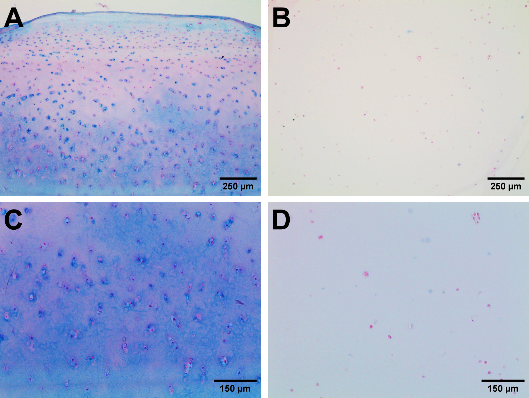
Figure 1. Articular cartilage tissue (A, C) and alginate-based hydrogel (B, D) stained following Alcian Blue & Nuclear Fast Red staining as per above indications. Sulfated polysaccharides are stained in blue, nuclei are stained in red, and extracellular maxtrix components are stained in pale pink. As can be appreciated in the images, no blue staining is detected in the alginate hydrogel and only nuclei are stained in red, whilst a strong blue signal is detected in the ECM of cartilage tissue.
| Number | Category | Product | Amount |
|---|