INTRODUCTION
Hematoxylin-Eosin stain (H&E stain) is one of the main tissue stains used in histology. It is the most widely used stain in medical diagnosis and is often the gold standard in histopathology. H&E is the combination of two histological stains: hematoxylin and eosin. Nuclei stained with hematoxylin display a purplish-blue colour, whilst the extracellular matrix and cytoplasmic components appear stained in pink by eosin. Other cellular and tissular structures can display different shades, hues, and combinations of these colors after staining. This stain combination was first introduced in 1877 by N. Wissozky at the Kasan Imperial University of Russia.
The H&E staining procedure is the main used stain in histology because it can be performed quickly, has a low cost, and achieves a tissue staining pattern which reveals a considerable amount of microscopic anatomy; helping to easily discriminate and evaluate different tissues. For these reasons, H&E staining is an excellent tool for the evaluation of the proper differentiation and degree of maturity of tissue-engineered constructs.
MATERIALS
- Xylene
- 100º ethanol
- 95º ethanol
- 70º ethanol
- 70º acid ethanol
- Hydrochloric acid (HCl) 37%
- H2O, distilled
- Harris Hematoxylin
- 0.2% Eosin Y (aqueous)
- Glass coverslips
- Mounting medium (DPX or similar)
EQUIPMENT
- Chemical fume hood
- Microscope
- Coplin jars
- Tweezers
METHODS
Follow institutional and local guidelines for the safe handling and disposal of chemical reagents. Prepare 95º and 70º ethanol from 100º ethanol and distilled water. Prepare 70º acid ethanol by adding 1 mL 37 % HCl to 99 mL 70º ethanol. Harris Hematoxylin and Eosin Y solutions can be purchased in a ready-to-use format.
Start with formalin-fixed paraffin-embedded histological sections mounted onto microscopy glass slides. For performing the H&E stain, follow the steps listed below:
- Xylene, 10 min
- Xylene, 10 min
- 100º ethanol, 5 min
- 100º ethanol, 5 min
- 95º ethanol, 5 min
- 70º ethanol, 5 min
- H2O distilled, 5 min
- Harris Hematoxylin, 5 min
- Running tap water, 5 min
- 70º acid ethanol, 5 sec
- H2O distilled, 2 min
- 0.2% Eosin Y, 2 min
- 70º ethanol, 15 sec
- 95º ethanol, 30 sec
- 100º ethanol, 3 min
- 100º ethanol, 3min
- Xylene, 5 min
- Xylene, 5 min
- Mount glass coverslips using DPX or similar
- Leave mounted histological preparations set overnight in horizontal position
- Visualize under brightfield microscopy
RESULTS
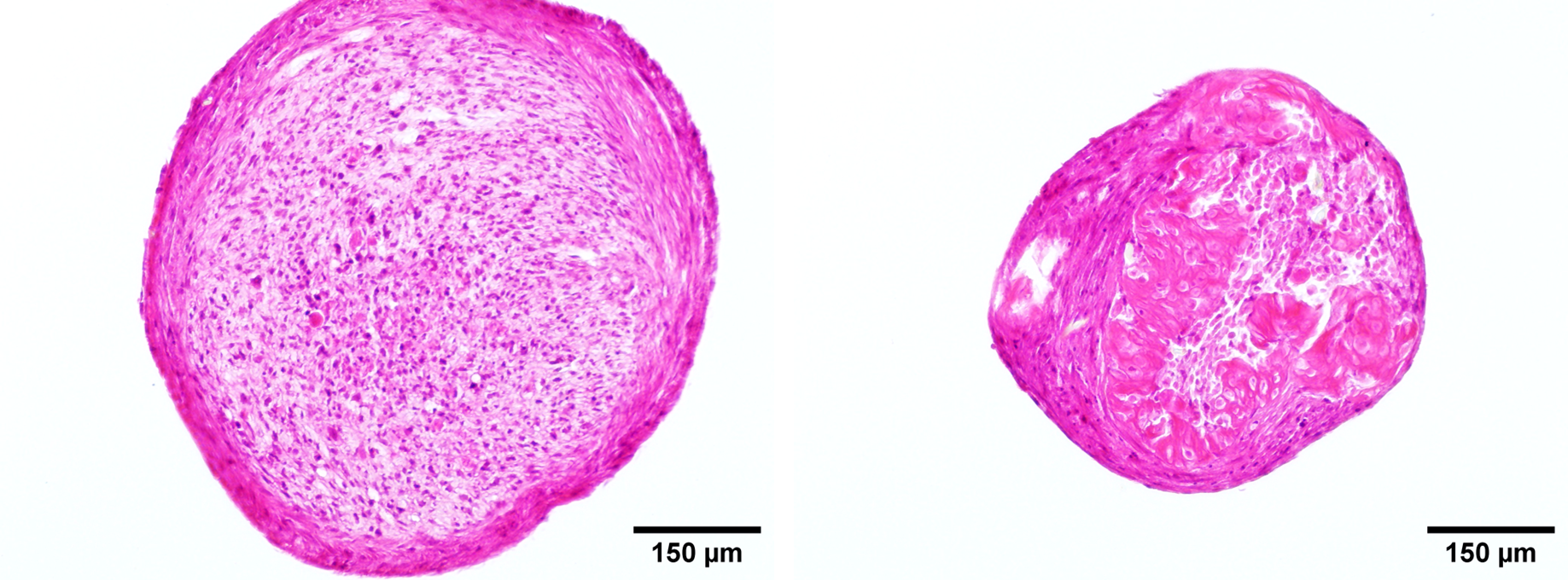
Following the protocol described above, staining of cellular and extracellular structures within tissue engineered constructs can be achieved. Nuclei will display a purplish-blue colour, whilst the extracellular matrix and cytoplasmic components appear stained in pink.
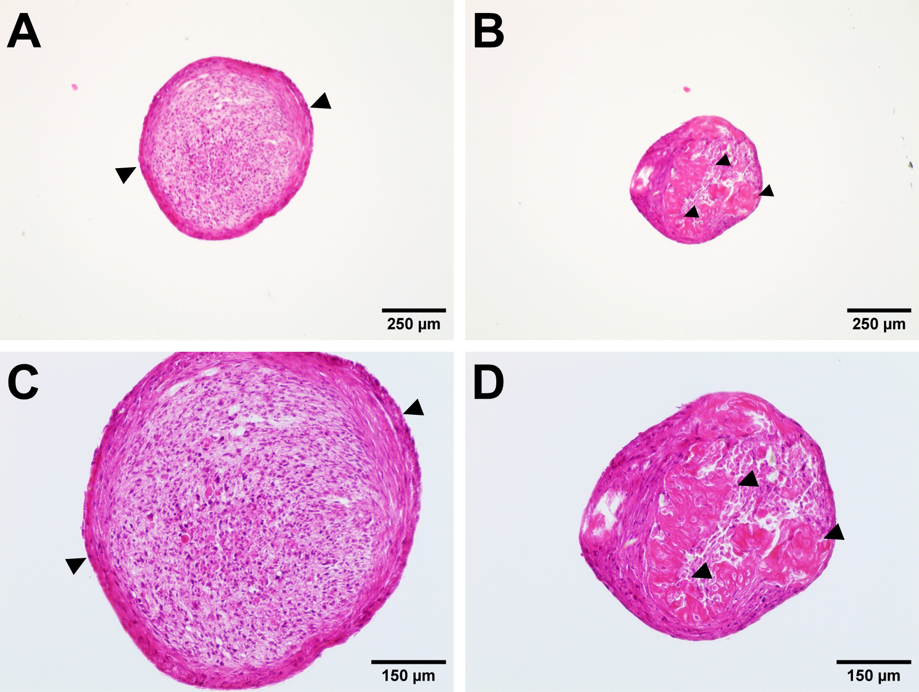
Figure 1. H&E staining of a mesenchymal stem cell (MSC) pellet (A, C) and a cell-laden hydrogel (B, D) cultured for 21 days in chondrogenic differentiation medium. A dense extracellular matrix capsule (arrowheads) stained with eosin can be appreciated in the periphery of the MSC pellet, surrounding numerous cells with pink cytoplasms and purple nuclei. An immature bone-like structure (arrowheads) stained with eosin can be appreciated inside of the MSC-laden hydrogel.
| Number | Category | Product | Amount |
|---|