
INTRODUCTION
Hydrogels are at the forefront of 3D bioprinting and tissue engineering. However, the wide variety of choices available, and the diverse physicochemical nature of the biomaterials comprising the hydrogels, complicate the selection of the most adequate formulation for each specific application. The purpose of this method is to evaluate the chondropermissive nature of hydrogel biomaterials for their further use as cell-laden hydrogels in cartilage 3D bioprinting and or tissue engineering. This method is based on the chondrogenic differentiation of mesenchymal stem cell pellets, described elsewhere.
MATERIALS
- Mesenchymal stem cells (MSCs); cryopreserved
- Dulbecco’s Modified Eagle’s Medium – Low glucose (DMEM-LG)
- Dulbecco’s Modified Eagle’s Medium – High glucose (DMEM-HG)
- Fetal bovine serum (FBS)
- Penicillin/Streptomycin solution (100x) (P/S)
- Phosphate buffered saline (PBS)
- Trypsin/EDTA solution (T/E)
- Trypan blue solution
- Tissue culture flasks (75 & 175 cm2)
- 15 mL conical polypropylene tubes
- 50 mL conical polypropylene tubes
- Serological pipettes (5, 10, 25 mL)
- Micropipette tips (10, 200, 1000 uL)
- Ascorbic acid 2-phosphate sesquimagnesium salt (5 mg/mL in dH20) (100x)
- L-proline (4 mg/mL in DMEM-HG) (100x)
- Insulin-Transferrin-Selenium supplement (ITS) (100x)
- Dexamethasone (10e-4 M in DMEM-HG) (1000X)
- Recombinant human TGFB3 (rhTGFB3)
- 4% paraformaldehyde in PBS
- 70% Ethanol
EQUIPMENT
- Biological safety cabinet
- Thermostatic water bath
- CO2 incubator
- Inverted microscope
- Centrifuge
- Automatic pipette controller
- Micropipettes
METHODS
Culture MSCs at 5,000 cells/cm2 in tissue culture flasks filled with 5 mL of DMEM-LG 10% FBS 1% P/S per each 25 cm2 of culture surface, refreshing culture medium every other day. Typically, MSCs are expanded up to passage 3 – 4 before performing chondrogenic differentiation. For subculturing MSCs, once culture dishes are at 85% confluence, remove culture medium, wash twice with PBS (2.5 mL per each 25 cm2 of culture surface), add T/E solution to the culture flasks (0.5 mL per each 25 cm2 of culture surface), and incubate for 5 minutes at 37ºC and 5% CO2 under humidified athmosphere. Monitor cell detachment using an inverted microscope, and once cells completely detach, recover with DMEM-LG 10% FBS 1% P/S at a ratio 2:1 (medium:T/E). Centrifugate the cell suspension at 150 xg in 50 mL conical tubes, and resuspend the cell pellet in DMEM-LG 10% FBS 1% P/S at a suitable concentration for cell counting (1 – 2 x 10e6 cells/mL). Dilute the cell suspension 1/2 with trypan blue solution and count cells using a hematocytometer.
Suspend cells at 2.5 x 10e5 cells/mL of DMEM-HG 10% FBS 1% P/S, add 1 mL of cell suspension to 15 mL conical polypropylene tubes, and centrifugate at 400 xg for 5 minutes. Incubate the tubes overnight with the resulting cell pellet at 37ºC and 5% CO2 under humidified atmosphere, with loosened caps to allow for gas exchange. Prepare cell-laden hydrogels by suspending MSCs in PBS at 10x the desired cellular concentration in the hydrogels (2 – 20 x 10e6 cells/mL recommended), and mix in a 1:9 ratio with the hydrogel/s of choice. Transfer 25 uL of cell-laden hydrogel to the bottom of 15 mL conical polypropylene tubes, and add 1 mL of DMEM-HG 10% FBS 1% P/S to each tube. Incubate the tubes overnight at 37ºC and 5% CO2 under humidified atmosphere, with loosened caps to allow for gas exchange. If using cross-linkable hydrogels, perform the corresponding cross-linking step before the addition of culture medium.
After 24 hours of culture, replace culture medium with DMEM-HG 10% FBS 1% P/S, 1% ITS, 50 ug/mL ascorbic acid 2-phosphate, 40 ug/mL L-proline, 10e-7 M dexamethasone, and 10 ng/mL TGFB3. Refresh culture medium every other day. After 21 days of culture, wash the samples 3x with PBS, and fix for 30 minutes with 4% PFA. After fixation, wash 3x with PBS and transfer the samples to 70% ethanol. Process the samples following routine histopathological paraffin embedding and sectioning, and stain the paraffin embedded sample sections with standard Hematoxylin & Eosin and or Alcian Blue stains.
RESULTS
 Figure 1. Representative images showing a MSCs pellet (A), a MSC-laden collagen-based hydrogel (B) and a cell-laden alginate-based hydrogel (C), after 24 hours in culture.
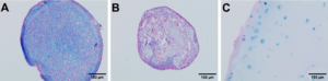
Figure 2. Representative images showing an Alcian Blue – Nuclear Fast Red stain of a MSCs pellet (A), a MSC-laden collagen-based hydrogel (B) and a cell-laden alginate-based hydrogel (C), after 21 days in chondrogenic differentiation medium. Chondrogenic differeniation can be inferred from the extracellular deposition of sulfated glycosaminoglycans, stained with the Alcian Blue dye. Nuclear Fast Red stain cell nuclei, cytoplasms and extracellular matrix in a red to pale pink colour.
| Number | Category | Product | Amount |
|---|