Introduction:
Injuries to the knee meniscus commonly lead to osteoarthritis. Current therapies for meniscus regeneration, including meniscectomies and scaffold implantation, do not achieve complete functional tissue regeneration. This has led to increased interest in cell and gene therapies and tissue engineering approaches to meniscus regeneration. The implantation of a biomimetic implant, incorporating cells, growth factors, and proteins derived from the extracellular matrix (ECM), represents a promising approach for regeneration of the functional meniscus.
Experimental Procedure:
Preparation of Chondrocytic Cells for Meniscus Regeneration:
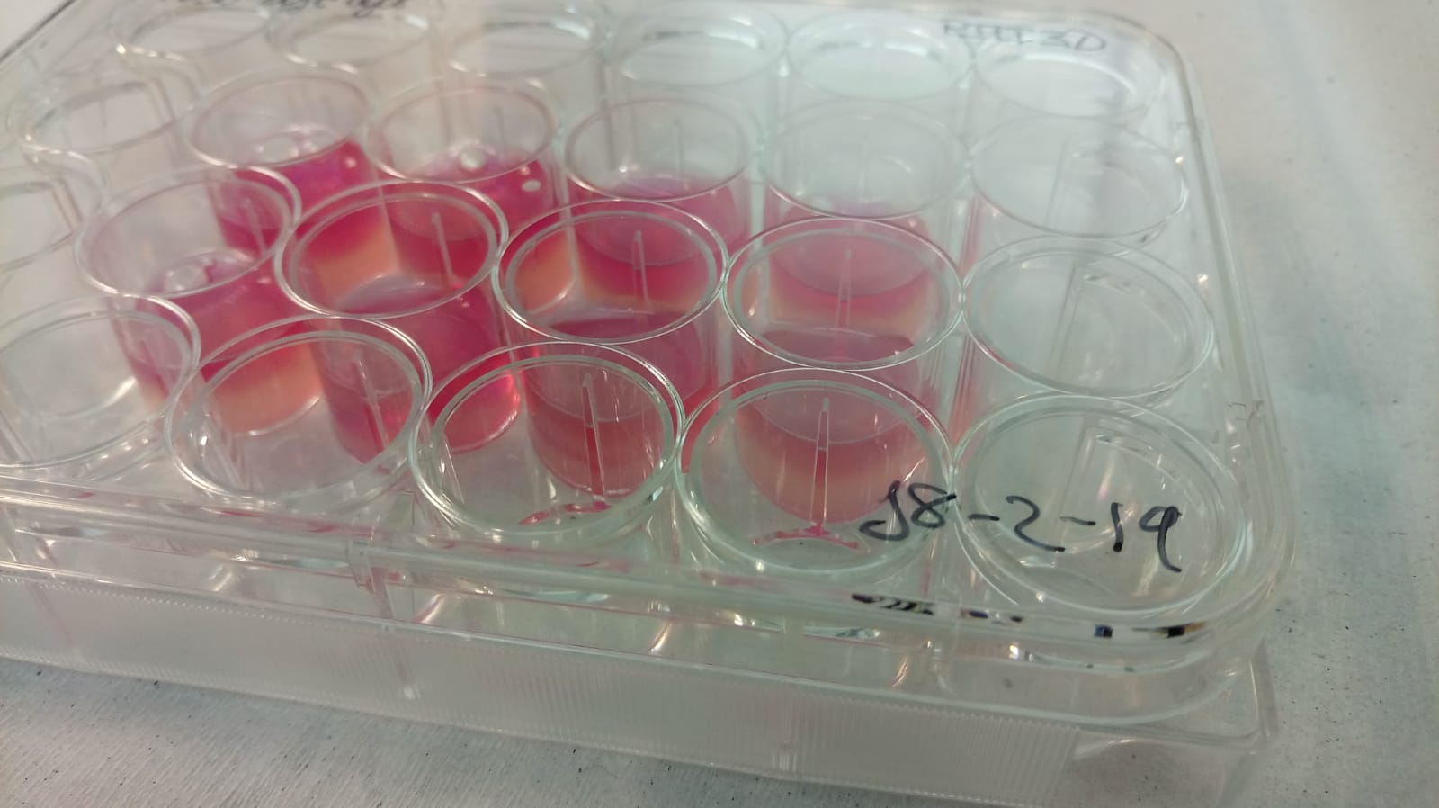
1. Examine Cell Viability to the Light Microscope. Cells maintained for a short number of passages and to be in high cell density are desired. Confluence = 85%.
2. Aspirate culture medium from the plate.
3. Wash with PBS using appropriate volume for each case. Usually this is about ½ of the volume used to feed the crop.
4. Add enzymatic dissociation medium (trypsin / acutase):
Plate Type Quantity to add per plate
24 well plate 0.250 ml
12 well plate 0.5 ml
6 well plate 1 ml
10 cm dishes 3 ml
5. Incubate plate for 10 minutes at 37ºC and 5% CO2.
6. After 10 minutes of incubation, check by microscope that the cells have completely disposed of the culture plate. If cells are still adhered, it is advisable to increase the incubation time.
7. Resuspend cells in dissociation medium gently using a p1000 pipet. Take care not to produce bubbles due to the possibility of decreasing cell viability.
8. Transfer cells to a 15ml conical tube.
9. Neutralize the dissociation medium, adding regular culture medium to the conical tube in a 1: 1 ratio.
10. Centrifuge the cells for 5 ‘at 200g-1500rpm.
11. Aspirate medium being careful not to scratch or remove the cell pellet adhering to the conical tube.
12. Resuspend pellet in 10ml of DMEM Glutamax cell culture medium (Gibco, Biosciences) + 10% FBS + 1% Pen / Strip.
13. Take aliquot of 10ul and mix with 10ul of Trypan Blue. Resuspend homogeneously and transfer to the Neubauer Chamber.
14. Proceed with the cell count applying the formula: No. of cells counted * Dilution factor applied (1: 2) * 104.
15. Take the volume necessary to obtain 2.5 million cells / mL.
Preparation of Hydrogel based on Nanocellulose and Alginate for Meniscus Regeneration:
1. Prepare stock solution of 2.5% Alginate and 8% Nanocellulose reduced by TEMPO technique.
2. Adjust the final volume of hydrogel diluted with Phosphate Buffer Saline (PBS) in a 1: 4 ratio.
3. Homogenize hydrogel by vortexing or centrifugation at 200g for 5 minutes.
4. Add DMEM GlutaMAX chondrogenic medium (Gibco, Biosciences, Ireland) supplemented with P / S, 100 μg / ml sodium pyruvate, 40 μg / ml L-proline, 50 μg / ml L-ascorbic acid 2-phosphate, 4.7 μg / ml linoleic acid, 1.5 mg / ml bovine serum albumin, 1X insulin-transferrin-selenium, 100 nM dexamethasone (Sigma-Aldrich, Ireland), and 10 ng / ml TGFβ3 (Prospec-Tany TechnoGene Ltd., Israel).
5. Incubate the biotint at 37ºC for 15 minutes to proceed with ionic cross-linking that facilitates the gelling of the hydrogel.
6. If cells are to be bioprinted directly, add 90% volume first and then the remaining 10% with cells after the incubation period.
Bioprinting of meniscus based on Alginate, Nanocellulose and Chondrocytes:
1. Add 10% cell volume to the gelled bioink.
2. Resuspend carefully, so as not to form too many bubbles, 10 times with a 1000ul pipette and cut tip.
3. Transfer biotin from conical tube to print cartridge.
4. Fill the cartridge with the largest possible volume in order to avoid the presence of an air phase inside the cartridge.
5. Install the cartridge in the bioprinter in tool T2.
6. Introduce PCL polycaprolactone thermoplastic filament into the extruder at a temperature of 190ºC.
7. Configure cylindrical figure with the bioprinter software with the following parameters:
to. Cylindrical .stl archive.
b. Diameter 25 mm.
c. Height 5mm.
d. Layer height: 0.35mm.
and. Pore width: 1.2mm.
F. Perimeters: 1.
g. Flow velocity: 1.5 mm / s.
The generated construct has to be incubated at 37ºC and 5% CO2 in measured DMEM GlutaMax for 21 days to obtain a mature graft. Proceed at medium change every 2-3 days.
| Number | Category | Product | Amount |
|---|---|---|---|
| 1 | - | Nanocellulose | 1 |
| 2 | - | Human Chondrocytes | 1 |
| 3 | - | DMEM Glutamax | 1 |
| 4 | Biomaterials | PLA standard-250 gram | 1 |
| 5 | - | Pen/Strep | 1 |
| 6 | - | REGEMAT 3D BioV1 Bioprinter | 1 |
One thought on “Cartilage tissue bioprinted models based on blendings of nanocellulose-alginate with embeded chondrocytes.”
excellent description!