Introduction
Fibrin is a degradable, resorbable biopolymer that naturally occurs in the wound healing process at the vascular level. It is formed from fibrinogen, a glycoprotein synthesised by the liver and found in blood plasma. When tissue damage occurs and results in bleeding, fibrinogen is converted into fibrin at the wound site, by means of the enzyme thrombin. Owing to its properties of biocompatibility, bio-resorbability, good cell interaction and growth promotion, as well as the tunability of its morphological and mechanical properties through the control of the precursor formulation, fibrin has already been investigated as an injectable gel scaffold and cell carrier in Tissue Engineering.
The present method aims at preparing a fibrin bioink for Cardiovascular Tissue Engineering (CTE), and assessing its cell viability.
Materials
The following materials were used:
- 1 and 10 mL syringes;
- 0.2 µL “button” filter;
- 15mL Falcon tubes;
- 75 cm^2 culturing flask;
- 48–well culturing plate;
- Extrusion conical tip (ϕ = 0.41 mm);
- NaCl;
- Fibrinogen powder (340 kDa, Sigma-Aldrich);
- Thrombin powder (Sigma-Aldrich);
- Bovine Serum Albumin (Sigma-Aldrich);
- Dulbecco’s Modified Eagle’s Medium (DMEM), high glucose, pyruvate (Gibco);
- Foetal Bovine Serum (FBS; Gibco);
- Penicillin (Lonza);
- Streptomycin (Lonza);
- Trypsin/EDTA;
- DBPS (Sigma-Aldrich);
- Live/dead kit (Biotium Inc, US):
- Calcein (green);
- Eth-D III (red);
- Human Umbilical Vein Endothelial Cells (HUVECs).
Equipment needed
- Biosafety cabinet Class II;
- Incubator;
- Centrifuge;
- Hot plate magnetic stirrer;
- Precision scale;
- Confocal microscope.
Method
Cell culturing
Cells are cultured in a 75 cm2culturing flask at 37◦C, 20% O2 and 5% CO2, in DMEM supplemented with 10% (v/v) FBS, and 1% of a mixture of 100 U/mL Penicillin and 100 µg/mL Streptomycin.
When the desired confluence is reached, cells are detached as follows:
- Aspirate medium and wash cells with DPBS.
- Incubate with Trypsin/EDTA at 37 ◦C for 3 min.
- Add FBS to the trypsin solution in a 2:1 volume ratio.
- Centrifuge the cellular suspension at 300G for 5 min.
- Discard the supernatant and resuspend the pellet in 1 mL of complete medium.
- Count cells.
Preparation of the fibrin gel precursors
- Prepare the fibrinogen and thrombin stock solutions.
- Fibrinogen: Firstly, prepare 10 mL of NaCl solution in ddH2O.(0.9% w/v). Heat the solution at 37◦C, and carefully layer the fibrinogen powder on top of the liquid surface to allow solubilisation, while under gentle stirring conditions. The process yields a hazy stock solution (10 mg/mL). Using a 10 mL syringe and a 0.2 µL “button” filter, filter the solution into a 15 mL Falcon tube. Store the solution at 4◦C for up to a week before use.
- Thrombin: The thrombin stock solution is also prepared at a concentration of 10 mg/mL, which in terms of activity equates to 454 U/mL. Dissolve the entire quantity of thrombin powder purchased in a solution of BSA in ddH2O (0.1% w/v). The solution can be stored in the fridge at 4◦C for up to one week before use.
- Dilute the stock solutions to the desired working concentration.
- Fibrinogen: dilute the stock solution in DMEM to a final fibrinogen concentration of 7.5 mg/mL.
- Thrombin: Dilute the stock solution to a final activity of 200 U/mL.
Gel cellularisation
To prepare 1mL of the final HUVEC–laden bioink, follow the procedure below.
- From the cell count, calculate the volume of cellular suspension (vC) needed to reach the desired cell density in the final bioink (∼ 10^6 cells/mL).
- Fill a 1mL syringe (S1) with the diluted fibrinogen solution previously prepared. The required fibrinogen volume is vF = (500µL −vc/2).
- Carefully inject the cell suspension volume vc in the fibrinogen–filled syringe, from the tip.
- Load a second 1mL syringe (S2) with a volume vM(= vF) of a mixture of BSA (0.1% v/v in ddH2O) and diluted thrombin solution previously prepared. The volume ratio between BSA and thrombin is 1:0.5.
- Equip S1 with one extrusion tip and gently extrude equal volumes (vW ≃ vF/3) of the HUVEC–laden fibrinogen gel in 3 different wells of a 48–well plate to have technical triplicates.
- Equip S2 with the extrusion tip and extrude a volume vW of the mixture in each well, to reach a fibrinogen:BSA:thrombin volume ratio of 1.5:1:0.5.
- Gently mix the compound in each well using a pipette tip.
- Top up each well with complete culturing medium.
Cell viability assay
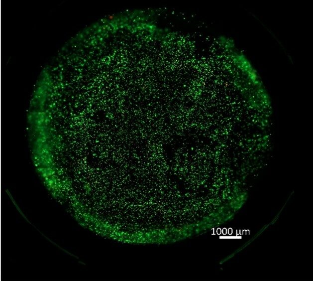
At the time of the first time point, discard the culturing medium from the wells, wash the constructs with DPBS, and incubate for 30 min at 37◦C, 5% CO2 with a solution of Calcein (1:2000 v/v in serum-free medium) and EthD-III (1:500 v/v in serum-free medium). Calcein is a fluorescent green dye that stains live cells, while Eth-D III is a red fluorescent dead cell staining dye.
After the analysis at the confocal microscope, put the constructs in the incubator and change the media every third day. Other time points can be assessed, to monitor the cell viability over time.
| Number | Category | Product | Amount |
|---|---|---|---|
| 1 | - | Human Umbilical Vein Endothelial Cells | 1 |
| 2 | - | Live/Dead kit [Calcein + Eth-D III] (Biotium Inc.) | 1 |
| 3 | - | DPBS (Sigma-Aldrich) | 1 |
| 4 | - | Trypsin/EDTA | 1 |
| 5 | - | Streptomycin | 1 |
| 6 | - | Penicillin | 1 |
| 7 | - | DMEM high glucose, pyruvate | 1 |
| 8 | - | Foetal Bovine Serum (FBS) | 1 |
| 9 | - | Bovine Serum Albumin | 1 |
| 10 | - | Thrombin powder (Sigma-Aldrich) | 1 |
| 11 | - | Fibrinogen powder (340 kDa; Sigma-Aldrich) | 1 |
| 12 | - | NaCl | 1 |
| 13 | - | Extrusion conical tip (ϕ = 0.41 mm) | 2 |
| 14 | - | 48–well culturing plate | 1 |
| 15 | - | 75 cm^2 culturing flask | 1 |
| 16 | - | 15mL Falcon tubes | 5 |
| 17 | - | 0.2 µL “button” filter | 1 |
| 18 | - | 1 mL syringes | 3 |
| 19 | - | 10 mL syringe | 1 |