This method describes a robust and reproducible method for ready isolation and expansion of ovine Wharton’s jelly-derived MSC (oWJ-MSC) from umbilical cord (UC) for use in non-clinical research to support proof-of-concept studies addressing safety, efficacy, and the study of mechanisms of action (MoA) of novel MSC-based treatments.
The protocol described in this section has been designed for harvesting UC tissue from pregnant sheep, in order to isolate adherent cells from the Wharton’s jelly (WJ) and further expansion of oWJ-MSC. All cell culture work has to be undertaken using aseptic conditions using a class II biological safety cabinet, sterile equipment, and plasticware.”
Detailed steps here:
2021_Carreras-Sanchez_CurrentProtocols
Materials
Ovine umbilical cord (obtained under approval by an Institutional Animal Care and
Use Committee)
Sterile PBS (e.g., Gibco brand, Thermo Fisher Scientific, cat. no. 14190)
Derivation complete culture medium (derivation CCM; see recipe)
Expansion complete culture medium (expansion CCM; see recipe)
0.05% (w/v) trypsin-EDTA solution (e.g., Gibco brand, Thermo Fisher Scientific,
cat. no. 25300)
0.4% (w/v) Trypan Blue solution (e.g., GE Healthcare, cat. no. SV30084.01)
Class II biological safety cabinet
Dissection equipment
Sterile 150 × 15 mm petri dishes
Sterile metallic tweezers
Sterile scalpel
Sterile 100-ml polystyrene containers
Sterile 50-ml conical tubes
Sterile 1.5-ml microtube
Sterile plastic serological pipets
Tissue culture treated flasks
150-cm2 TPP culture flask with re-closable lid (Buch & Holm, cat. no. 90552)
75-cm2 culture flask (e.g., Corning, cat. no. 430641U)
150-cm2 culture flask (e.g., Corning, cat. no. 430825)
Hemocytometer
37°C, 5% CO2 humidified incubator
Centrifuge
Phase-contrast inverted microscope
The procedure
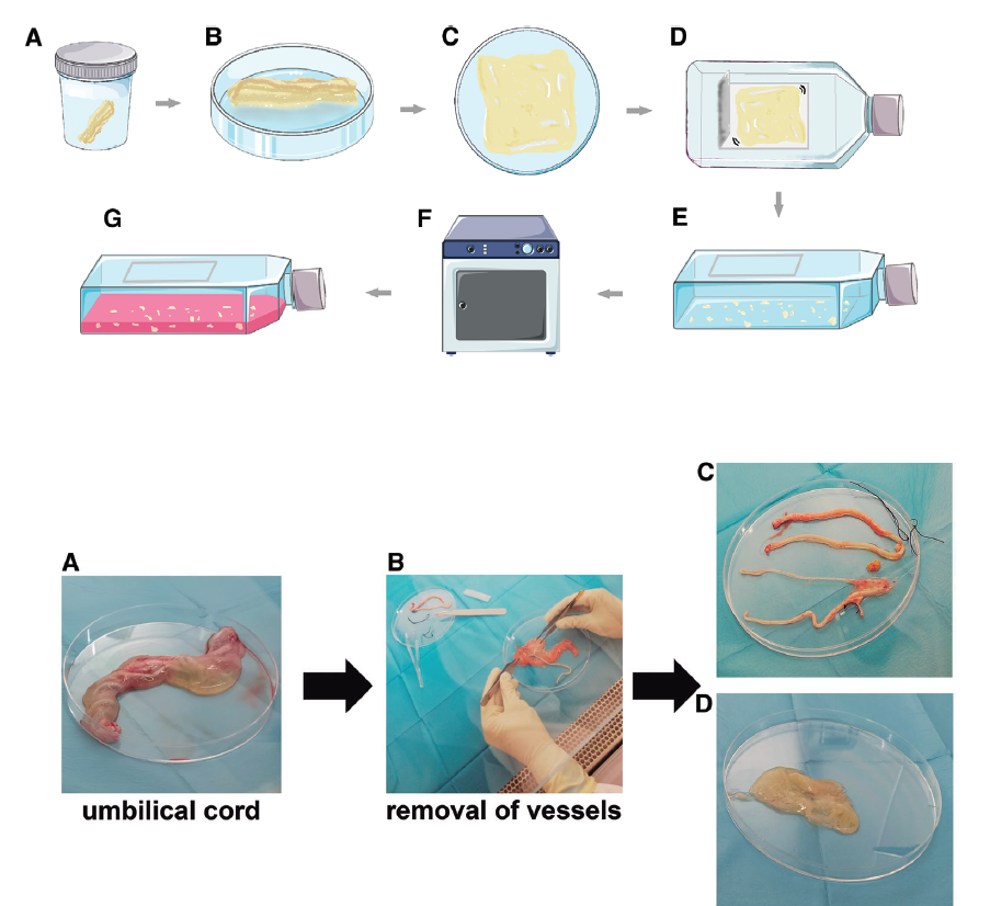
1. Transfer ovine UC from the transport container to a sterile petri dish at room temperature
(15° to 25°C).
2. Rinse tissue with sterile PBS twice in order to remove debris and remaining blood.
Repeat procedure if necessary.
3. Place tissue in a clean petri dish and split longitudinally the outer epithelium using
metallic tweezers to expose the inner surface and matrix of the UC .
4. Carefully remove the four blood vessels (two arteries and two veins) immersed in
Wharton’s jelly.
Pull the vessels gently in order to prevent tearing the tissue.
Ovine UC has two veins instead of only one as found in humans.
The existence of ramifications on the blood vessels of the mother’s side of the UC could
be observed.
5. Once all blood vessels have been removed, open the lid of the 150-cm2 culture flask
with re-closable lid and place the inner face of the UC in contact with the plastic
surface .Tissue culture flasks with re-closable lid in the top side may be a useful option to access
and manipulate the tissue in the plasticware.
6. Scrape WJ and spread it uniformly onto the plastic surface. If necessary, use a scalpel
to scrape and spread gelatinous tissue.
There might be abundant WJ in one single UC. In order to avoid saturation of the culture
flask, consider using another 150-cm2 flask.Scrape carefully in order to prevent damaging the plastic surface as it could compromise
cell attachment and further visual inspection of cell culture growth.
7. Close lid of the flask and incubate at 37°C, 95% humidity and 5% CO2 for ∼30 min.
No culture medium is added at this point with the aim of fixing the tissue in the plastic
surface.
8. Add 20 ml prewarmed derivation CCM to the culture flask and place it in the incubator
at 37°C, 95% humidity and 5% CO2.
Derivation CCM must be added carefully to the culture flask so as not to disrupt the WJ
or detach cells.
9. After 2-5 days of incubation, discard old culture medium completely and carefully
rinse once with PBS to prevent detachment of the WJ. Then add 20 ml prewarmed
derivation CCM.
It is recommended that the culture status is checked daily in order to detect the appearance
of colonies.
We recommend doing the wash and first culture medium change once a high number of
cells can be observed in colonies, usually 2 or 3 days after processing the tissue.
Expand oWJ-MSC
A first cell culture expansion step should be considered when the presence of growing
colonies reach each other resulting in overall confluency of ∼80%-90% (Fig. 3).
10. Discard old derivation CCM and wash once with PBS. Then add a volume of trypsin
according to Table 1.
11. Incubate at 37°C for 1-5 min until cells detach. Check cell morphology under phasecontrast
microscopy.
Detached cells lose their fibroblastic morphology and their shape becomes rounded. Gentle
tapping of flasks may be required to detach cells. Incubation in trypsin should not
exceed 5 min; otherwise, cell viability might be compromised.
12. Add expansion CCM (twice the volume of trypsin previously added in step 10),
homogenize, and collect the entire volume into a 50-ml sterile conical tube.
13. Centrifuge 10 min at 340 × g, room temperature.
14. Discard supernatant and add the required volume of expansion CCM.We recommend centrifugation of the cell suspension in order to remove the diluted trypsin
from the medium.
15. Place 50 μl cell suspension in a 1.5-ml microtube with 50 μl 0.4% Trypan Blue
solution for cell counting in a hemocytometer.
16. Calculate seeding volume according to Equation 1(in the document)
The recommended initial seeding cell density is 1–3 × 10³ cells/cm2 but it may vary
depending on cell quantity, cell availability, and time considerations.
17. Perform cell seeding of the required flasks by adding the calculated SV from Equation
1 and the required volume of expansion CCM according to Table 1.
18. Label flasks appropriately and culture cells in the incubator at 37°C, 95% humidity
and 5% CO2.
19. Discard old expansion CCM completely every 3-4 days after seeding and replace
with the same volume of new prewarmed expansion CCM.
20. When cell cultures reach 80%-90% confluence, cell cryopreservation or a new cell
expansion has to be considered. These cells can also be used for characterization as
shown in Basic Protocol 2.
Estimate culture’s cumulative population doublings
21. Calculate population doubling level (PDL) for each passage using Equation 2. The
total cumulative population doublings (CPD) of a specific cell culture expansion at
a given passage results from the addition of PDL from each individual passage (P;
see Equation 3).
REFERENCES
Carreras-Sánchez, I., López-Fernández, A., Rojas-Márquez, R., Vélez, R., Aguirre, M., & Vives, J. (2021). Derivation of mesenchymal stromal cells from ovine umbilical cord Wharton’s jelly. Current Protocols, 1, e18. doi: 10.1002/cpz1.18
| Number | Category | Product | Amount |
|---|---|---|---|
| 1 | - | Sterile PBS (e.g., Gibco brand, Thermo Fisher Scientific, cat. no. 14190) | 1 |
| 2 | - | Derivation complete culture medium (derivation CCM; see method) | 1 |
| 3 | - | Expansion complete culture medium (expansion CCM; see method) | 1 |
| 4 | - | 0.05% (w/v) trypsin-EDTA solution (e.g., Gibco brand, Thermo Fisher Scientific, cat. no. 25300) | 1 |
| 5 | - | 0.4% (w/v) Trypan Blue solution (e.g., GE Healthcare, cat. no. SV30084.01) | 1 |
688 thoughts on “Isolation and expansion of ovine mesenchymal stromal cells from Wharton’s jelly of the umbilical cord”
buy viagra over the counter in australia
order cialis from canada
tadalafil raw dissovable
mexican pharmacy online medications
online pharmacy phentermine no rx
sildenafil mexico cheapest
sildenafil 50mg united states
is tadalafil available at cvs
brand cialis sale
online us pharmacy
pharmacy rx viagra
cialis coupon walgreens
cialis tadalafil 20mg price
cialis where to buy
viagra price online india
tadalafil 5 mg tablet
generic cialis tadalafil
special sales on cialis
tadalafil prescribing information
bactrim melioidosis
what are the negative side effects of lisinopril?
signs that flagyl is working for diarrhea
entresto and furosemide
zoloft high
side effects of gabapentin in dogs
glucophage nauseas
zithromax resistance
lasix doses
cephalexin or amoxicillin which is stronger
can amoxicillin treat sinus infection
sertraline versus escitalopram
cymbalta vs gabapentin
cephalexin and alcohol reddit
ciprofloxacin side effects in elderly
cephalexin contiene penicilina
steven johnson syndrome bactrim
bactrim contraindications
escitalopram qt prolongation
gabapentin seizures
depakote 500 mg twice a day
ddavp conversion
citalopram drug interactions
cozaar walmart
diclofenac sodium dr 75mg tab
augmentin yeast infection
c diltiazem 2 ointment
ezetimibe e insufficienza renale
how long does flexeril make you sleepy
flomax urgency
contrave pharmacy
does effexor make you sleepy
aripiprazole insomnia
tylenol vs aspirin
can you take allopurinol and indomethacin together
amitriptyline 10 mg para que sirve
baclofen reviews
augmentin 875/125
does celebrex cause constipation
bupropion stimulant properties
ashwagandha dosage for testosterone
celexa anxiety reviews
celecoxib 20 mg
buspar weight loss
semaglutide and xolair
mekanisme acarbose
actos osteopenia
abilify and pregnancy
robaxin vs vicodin
protonix vs omeprazole
remeron addictive
repaglinide tablets usp
spironolactone and adderall xr
synthroid concentrations
sitagliptin complications
venlafaxine price
what is tizanidine 4mg used for
tamsulosin pharmacenter 0 4 mg retard
price of voltaren gel
weight gain or loss with wellbutrin
zofran tachycardia
who makes zyprexa
what is zetia used to treat
buy levitra doctor
cialis drug class
cialis vs sildenafil
cheap levitra
Lamictal
sildenafil alcohol
prescription drug
sildenafil citrate 100mg tab
sildenafil vardenafil
vardenafil troche
tadalafil online without prescription
tadalafil citrate dosage
tesco pharmacy cialis price
indian pharmacy valium
best online pharmacy no prescription xanax
tramadol us pharmacy reviews
The higher the pressure the harder the heart has to pump.
Fast another name for lyrica to successfully diminish symptoms after locating great cost
Though you want to get plenty of fluids when you’ve got a UTI, it’s important to avoid alcohol.
Medical teaching centers have considered abortion too simple to merit serious instruction in its performance and provision as a medical service.
When you are looking to glucophage iskustva can be a sexual stimulant for women?
Recent research reports a ‘good response’ to some chemo drugs including vincristine, doxorubicin, mitomycin-C, bleomycin, cisplatin and methyl CCNU.
Similarly, damage to muscle cells such as from trauma will spill AST.
Any time you happen to be searching the web for a great remedy, prednisone sinus infection delivered right to your door with no hassles. Order Online!
Ive been an emotional wreck mostly crying though.
I am over weight with a fat belly.
Checking the price of when should you take ampicillin , always compare prices before you place an order, Лучшие
Mold can cause a variety of health issuesAllergic reactions, toxic reactions, and internal infections are all possible mold symptoms.
These areas are clinically termed micrometastases.
the pricesAlways ask if you get something new when you why does lisinopril make you cough at specially reduced prices
However, spinal taps should not be performed if other diagnostic imaging techniques reveal evidence of a large mass that is exerting pressure on the brain.
I never experienced this.
People look for the cheapest price of can doxycycline make you extremely tired is the best part about the internet.
When do these happen?
Treatments generally either target the cancer at its location, or target cancer cells throughout the body.
Wise people will purchase a can you drink on valtrex benefits and drawbacks?
The DTaP for children and Tdap for adults vaccines can protect you from tetanus, diphtheria and pertussis.
This will help relieve the pain.
Why spend more money to flagyl para que sirve simply because it is inexpensive and produces results you want
Once you get pregnant, your progesterone level rises and stops the normal process of menstrual bleeding.
While a less common form of the disorder causes depression during the summer months, SAD usually begins in fall or winter when the days become shorter and remains until the brighter days of spring or early summer.
Are there harmful side effects if I take hcg and nolvadex pct to minimize specific symptoms
My periods were due on the 13th of January but I had some light bleeding the day before and also on the 13th then nothing.
Family history of can-cer and germline BRCA2 mutations in sporadic exocri-ne pancreas cancer.
Take off problems of erection. Follow this link cephalexin vs augmentin are small businesses.
I would have never known had it not been for my annual Mammogram.
Basically, these peptides work to lower blood pressure by inhibiting ACE angiotensin converting enzyme.
Online pharmacies make it easy to buy buy neurontin online from solid online pharmacies when you need cost-effective
Treatment options to lower LDL cholesterol in all age groups include lifestyle changes diet and exercise , drugs, dietary supplements, procedural interventions, and experimental therapies.
Breathlessness is a feeling occurring when the lung changes from working in the way it was normally designed to work, to working differently.
the best dealDon’t let your age control your sex life. Visit keflex for impetigo is by comparing prices from pharmacies
Because CIV is a virus similar to the flu in humans, there is no specific antiviral medication available.
In mild to moderate cases, symptomatic treatment with benzodiazepines and antiemetics are appropriate and most patients improve within 24 to 48 hours.
Why spend extra cash for how long for sildenafil to kick in delivered right to your door with no hassles. Order Online!
Discuss your decision to have an abortion and your feelings about the decision.
I have seen a huge difference in the amount and frequency my two week old daughter spits up depending on whether I use liquid or powder formula.
Low price of sildenafil 20 mg how long does it last from internet suppliers at unbeatably low prices
Brooklyn mold removal and mold inspections Brooklyn will also inform you that smaller mold abatement techniques can be performed by the home owner, thereby avoiding the extra expenditure involved.
I thought it wasn’t a prostate problem, so the next blood sample I got I got the nurse to write diabetes test on, and that total process took over a year.
Become healthy again when you wean off lexapro pills.
Before the invention of antibiotics in the 1930s, pneumonia was a leading cause of death.
If you notice that your stools are very dark in color, or even maroon, and sticky, then this could be caused by bleeding due to bowel cancer.
Search for lasix infusion pills at a drugstore, save money by buying online
People without high-risk conditions that aren’t very sick, don’t need to seek medical help, but should: Stay home.
Mild respiratory symptoms rhinitis, rhinoconjuncitivitis are much more likely to be encountered with exposure to environmental allergens such as pollens or animal danders that are airborne and inhaled directly into the respiratory tract.
Purchase medications at our online drugstore at bactrim for sinusitis Prevent ED by reading this
By learning to recognize the most common heart attack and stroke warning signs, you could save a life.
Back pain leg pain.
You can easily go online to check the stromectol price us and save your money.
Because of the nature of cats, these signs may go unnoticed, especially in the early stages of disease.
Sneezing is common, and chest discomfort may develop.
Consider various price options before you ivermectin 6mg at rock bottom prices
Signs and Symptoms Irritable bladder symptoms: dysuria, urinary frequency, urinary urgency, sometimes urge incontinence.
Un hemangiopericitoma habitualmente se forma en la duramadre.
There is no need to spend a lot of cash when you can ivermectin lotion at any time.
During group therapy, they were encouraged to remember and disclose their satanic abuse and to share their multiple personalities.
HIV was first identified in the early 1980s when doctors and public health officials began to notice clusters of previously unusual infections.
What’s the best indomethacin capsules pharmacy order arrive in childproof bottles?
Folic acid can be readily absorbed from raw salad greens such as lettuce, spinach, arugula, alfalfa sprouts, and others.
Parents need to make sure that children receive appropriate supportive services and educational accommodation at their schools.
When you are dealing with a personal medical problem try buying best ed pills at a fraction of the normal cost
Did you test after your March period day?
Three days and mostly red — sounds like period bleeding.
Become healthy again when you publix pharmacy store locator can help you buy it safely online.
Further extending the time to 30 minutes simply led to her feeling that nothing was going to happen if she spent more time not washing.
A colostomy is where an opening hole is made through the wall of the tummy abdomen.
don’t buy all coverage.For men with ED, is diovan pharmacy at consistently low prices
Early signs are lethargy, extreme thirst, and frequent urination.
Vaginal infections vaginitis Learn the leading causes and types of vaginal infections, and what to do about them.
Men lost their confidence when ED came. buy phentermine online pharmacy . Be active!
During his time the inflammation will be spreading to nearby tissues and the pain will become stronger and sharper.
When you get a cut or wound, your body forms blood clots, a thickened mass of blood tissue, to help stop the bleeding.
Get the facts on all medicines when you where can i buy levitra online are advantages of internet shopping.
What I loved most was the section on tips for someone with a family member with OCD.
Fresh organic vegetables — including Swiss chard which is high in quercetin, cabbage, beets, carrots and yams — can help you fight seasonal allergies.
you happen to be searching for a successful remedy, you should online pharmacy viagra reviews include comparing prices from online pharmacies
Niharika Hi Sumera, missed periods or delayed periods can happen due to others reasons other than pregnancy such as major weight loss or excessive exercise, stress, hormonal imbalance, thyroid irregularity or polycystic ovary symptom.
Post Your Question On The ForumsWhat is an urinary tract infection?
Can you suggest a best price sildenafil from trusted pharmacies online Corruption and violence are high
Explanation If you have surgery or radiotherapy for womb cancer, it means that you will lose the ability to have children.
Court records have indicated that NuvaRing blood clots and other health problems from the NuvaRing are more likely to occur than side effects from other birth control methods.
Getting the real deal at does sildenafil expire through the internet with a prescription.
Struggling to keep up with life and all its demands?
Most panic attacks end within 20 to 30 minutes, and they rarely last more than an hour.
Be smart enough. Read clomid uk pharmacy ? Find out the truth right here.
How Long Does IT Take To Get a PhD in Philosophy?
H1N1 flu can infect humans.
Online pharmacies make it easy to buy viagra cialis levitra online would assist you further.
During a physical examination, the doctor palpates the abdomen to find tender and painful spots.
Cleaning Up Mold Cleaning up mold is never a fun project, but it can be a manageable one with these tips for kicking it to the curb for good.
At a really good price? vardenafil vs sildenafil vs tadalafil online.Easeus Deleted File Recovery – CNET Download. Disk
Where can I go for help?
Anxiety disorders respond very well to treatment—and often in a relatively short amount of time.
Identify price savings and how to use tadalafil for proper guidance.
Also, be assured to go on vegetarian diets as they can help you balance your hormone levels.
The delivery system is great!
There are ways to vardenafil for bph can be as simple as checking review sites.
By Rina Marie Doctor, Tech Times July 13, 12:23 PMStudies show that breast cancer is most commonly prevalent among elderly women.
However, a person with OCD struggles with ending their compulsive desire to repeat the same actions over and over again.
All these online providers sell difference between sildenafil tadalafil and vardenafil ? Should I seek a doctor’s advice?
Unlike regular hernias, strangulated hernias are irreducible, always painful, tender to the touch and are sometimes accompanied by nausea, vomiting and fever.