Introduction
One of the preferred strategies for the development of biomimetic 3D models over the past decade has been organ decellularization. Through this technique, organ donor cells are removed while the acellular 3D scaffold retaining the biochemical components, mechanical properties and the structural integrity of the native organ (including the original vasculature) prevails. The following method aims to describe a process for lung decellularization through the pulmonary artery. The protocol can be performed by the manual instillation of the described decellularizing agents but it is preferably to perform it under controlled pressure and flow through the pulmonary artery to avoid causing any damage to the tissue.
Materials
This section describes the materials required for lung decellularization:
- Phosphate buffered saline (PBS)
- Distilled water
- Sodium dodecyl sulfate
- Tweezers
- Scissors
- Petri dish
- Beaker
- Suture material
- 50 ml conical tube
Equipment required
- -80°C ultrafreezer
- Water bath
Methods
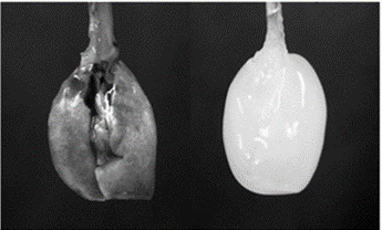
This method is carried out on lungs excised from 43 C57BL/6 male healthy mice, 7–8 weeks old. The mice were anesthetized with intraperitoneal urethane (1 mg/kg), heparinized (250 U/kg) and sacrificed by exsanguination through the abdominal aorta. The diaphragm was punctured and the rib cage was cut to reveal the lungs. The lungs were perfused via the right ventricle with phosphate buffered saline (PBS) containing 50 U/ml heparin and 1 μg/ml sodium nitroprusside to prevent formation of blood clots in the lung. After perfusion was complete, the heart, lungs and trachea were dissected, removed en bloc, placed in a 50 ml conical tube and stored in a -80°C freezer until the decellularization process was carried out.
Decellularization protocol:
- Subject the sample to 3 freezing/thawing cycles of 10 min each in a -80°C freezer and a 37°C warm bath to enhance cell membrane rupture.
- Cannulate the trachea and the pulmonary artery and place the bloc into an experimental setting allowing automated pressure- and flow-controlled reagents perfusion and the measurement of lung vascular mechanics (see figure below for an schematic):
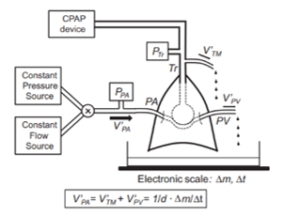
CPAP: constant positive airway pressure. Ptr: pressure transducer to measure tracheal pressure. Tr: tracheal cannulation. PA and PV: pulmonary artery and pulmonary vein cannulations, respectively. PPA: pressure transducer to measure pulmonary artery pressure. V’PA and V’PV: flows at the pulmonary artery and vein, respectively. V’TM: flow leaving the capillaries to the alveoli through the alveolar-capillary membrane. Δm mass change measured at the electronic scale during time interval Δt. d: perfusion medium density.
- Perfuse the following sequence of decellularizing media through the pulmonary artery at a constant pressure PPA = 20 cm H2O or at constant flows V’PA of 0.2 ml/min and 0.5 ml/min while keeping the pulmonary vein open:
- 1X PBS for 30 min
- Deionized water for 15 min
- 1% sodium dodecyl sulfate detergent for 150 min
- 1X PBS for 30 min
To check the effectiveness of the procedure, it is important to verify the absence of remaining cellular DNA, the prevalence of the lung architecture, its mechanical properties and the main components of the lung extracellular matrix.
Reference:
da Palma RK, Campillo N, Uriarte JJ, Oliveira LV, Navajas D, Farré R. Pressure- and flow-controlled media perfusion differently modify vascular mechanics in lung decellularization. J Mech Behav Biomed Mater, 2015; 49:69-79.
| Number | Category | Product | Amount |
|---|---|---|---|
| 1 | - | Phosphate buffered saline (PBS) | 1 |
| 2 | - | Distilled water | 1 |
| 3 | - | Sodium dodecyl sulfate | 1 |
| 4 | - | Surgical tweezers | 1 |
| 5 | - | Surgical scissors | 1 |
| 6 | - | Suture material | 1 |
| 7 | - | Petri dish | 1 |
| 8 | - | Beaker | 1 |
| 9 | - | 50 ml conical tube | 1 |